On June 25, 2026, a five-person lab called Aleph Neuro released a video showing the highest-resolution 3D images of a living human brain ever captured from outside an intact skull. A follow-up two days later told the story of how the team got there. The numbers matter only because the claim is large, and large claims usually get pushed back on fast. This one mostly did not.
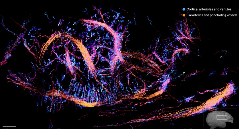
The images show blood. The reconstructions map the brain's vasculature in fine detail, down to pial arteries and small arterioles, and Aleph says the volumetric resolution is roughly 100 times finer than that of a comparable CT scan. That number is the lab's own, and it is the part to check as others work with the data.
Why is blood a reasonable proxy
The method rests on neurovascular coupling. When a patch of cortex becomes active, the blood flow to it rises within a second or two. Measuring where and when blood moves gives you an indirect read on where the brain is working. This is the same principle fMRI uses. Aleph's bet is that you can get a useful version of that signal with ultrasound instead of a room-sized magnet.
Getting an ultrasound to say anything accurate through the skull is the hard part. Bone scatters and distorts the sound, which is why ultrasound has never been the tool of choice for the brain. Aleph's approach borrows a technique called ultrasound localisation microscopy. The patient receives an injection of microbubbles, gas-filled spheres a few micrometres across that are already approved as a contrast agent. Each bubble reflects sound strongly, and because the bubbles are sparse, the system can pin down the centre of each one to a precision far below the ultrasound wavelength. Track those points across thousands of frames, and you recover the vessels they flowed through, in three dimensions.
Co-founder Lev Chizhov prefers the raw version to the polished renders. It "doesn't look as fancy as the final image," he says, "but you can feel it: you see the individual microbubbles flowing through the brain."
The hardware underneath is Butterfly Network's Ultrasound-on-Chip, which replaces the usual piezoelectric crystal with a semiconductor, making a full ultrasound system small and cheap. Butterfly named Aleph an embedded partner in a release timed to the launch.

Set against the alternatives, the position is clear without ranking them. EEG and MEG read the brain from the scalp, but they are badly blurred because the signal spreads on its way out. fMRI and CT are sharp but immobile and expensive. Electrodes are sharp and local but require surgery. Aleph is trying to buy a chunk of the resolution of the big machines at the size and cost of a handheld probe. This version still leans on injected contrast. The stated direction is contrast-free imaging, reading the weak signal from red blood cells themselves, using better hardware and machine learning trained on what the lab says is the largest neurovascular ultrasound dataset in existence.
The release
Luckily for us, the team decided to open-source their project. The pipeline and the data are on GitHub under an open licence. Not a paper describing the method, and not a gallery of finished renders, but the reconstruction code and the raw bubble tracks. That matters for research because stroke, Alzheimer's, and traumatic brain injury all leave vascular signatures that current scanners can miss. It also had a second effect, which the lab may not have planned for. It lets other people make the images.
How the visuals were actually made
The renders that circulated the announcement across the internet were not strictly scientific reconstructions. A few weeks before launch, Aleph handed its scan data to the visual artist Sohrob at frustum_studio. The input was about as plain as data gets: "raw .csv files with hundreds of thousands of points," he says, each row holding "the xyz position of one bubble inside the brain (captured 222 times/second) and a track number linking it to that single bubble's full path." The brain in those files was Marley Xiong's, the CEO, who volunteered as the subject.
From there, it became a visual-effects problem. Sohrob took the CSVs into Houdini and "used Claude code and the Houdini MCP to port it into SideFX, then built my own toolset on top so I had total control of the look and feel." He turned the tracks into motion fields and performed simulations through them, one particle pass that "made the brain feel like a tree growing," and a pyro pass, the tool built for smoke and fire, that "pushed a smoky haze through the brain's vasculature."
The quality became its own problem. "Most of our renders of the brain were too beautiful to be used," Marley Xiong said, "and would make people think it was fake."
Sohrob was not the only one. Releasing the tracks, rather than the finished pictures, turned one dataset into many. Eric Porres took the same public data and built a 3D explorer you can move through in a browser, allowing you to "make some interesting visualisations with your data set indeed." Others asked for files to 3D-print, and Aleph handed over track data and pointed them to tools that could render it. A single scan became a scientific reconstruction, a set of cinematic renders, a browser explorer, and a physical print because the underlying points were free to use.
The team and the reason they are doing this
Aleph is a group of five people who describe themselves as renegade physicists and engineers: Marley Xiong, who runs it, along with Raffi Hotter, Lev Chizhov, Thomas Ribeiro, and Jeff Brown. None of them was ultrasound people to begin with. "Telepathy as a problem feels physics-complete: making new brain interfaces requires knowing all the laws of nature," Xiong wrote of the starting point. Xiong and Hotter left software for Boston and, in Xiong's words, "taught ourselves physics, ultrasound, electromagnetism by auditing classes and reading textbooks."
The lab that produced these images was a house. The team "bought a real human skull and built a lab at home," using tofu as a tissue phantom, building a jig to measure attenuation, making their own acoustic lenses out of Orbeez, and, by Xiong's count, "27 gallons of deionised water." The scrappiness ran to haircuts. Chizhov described an "improvised last-minute fade haircut for better signal quality," because "sometimes that's just what you have to do to see inside your brain." Even the name was a near-miss. "Aleph used to be called Alegria," Hotter said, "but people would misread it and call us the Algerian Neurotechnology Company."
The imaging is a means, not the end. What they are after is a brain interface that moves what they call latents, the raw content of a thought before it is flattened into words. They point to work like MindEye, which reconstructs images from fMRI recordings, and argue that a palm-sized ultrasound device could one day reach comparable decoding without the machine around it.
We believe telepathy will be as important and as natural as words and hands.
That is a long way from a vascular map, and they know it.
What is still a claim
The reception was warm across the medical-device press and the neurotech community, with coverage from MassDevice and an endorsement from Butterfly's chief executive, Joseph DeVivo. This enthusiasm is earned. The images are striking.
There are some caveats, too. Millimetre-scale vascular resolution is coarse compared with the single neurons that carry out computation. Mapping blood flow is not decoding thought, and the gap between the two is the problem. The early doubt that ultrasound could resolve anything through an adult skull was answered by the results, but the distance from here to a working interface is the part still to be built.
None of that subtracts from what was shown. A small team imaged the living brain through the skull at a resolution that did not look available at this size and cost, and then let everyone else pick up the data. For a field that usually moves behind closed doors and inside large machines, that is the part to watch.
References
- Announcement thread: x.com/alephneuro
- The team's story: x.com/alephneuro
- Technical write-up: alephneuro.com/blog/ultrasound-brain
- Aleph Neuro: alephneuro.com
- Code and data: github.com/alephneuro/microbubbles
- Sohrob on the visuals: x.com/sohrobxyz
- Eric Porres interactive: porres.com/alephbrain
- Butterfly Network release: businesswire.com
- MassDevice: massdevice.com